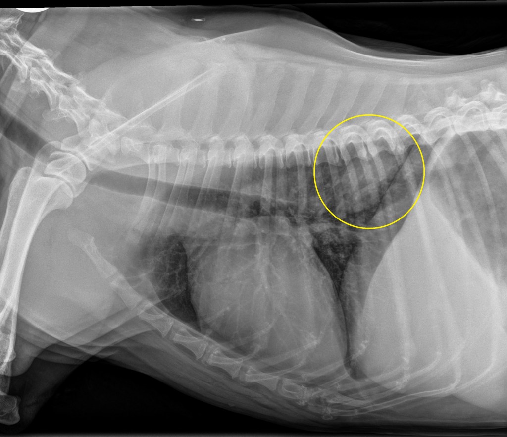

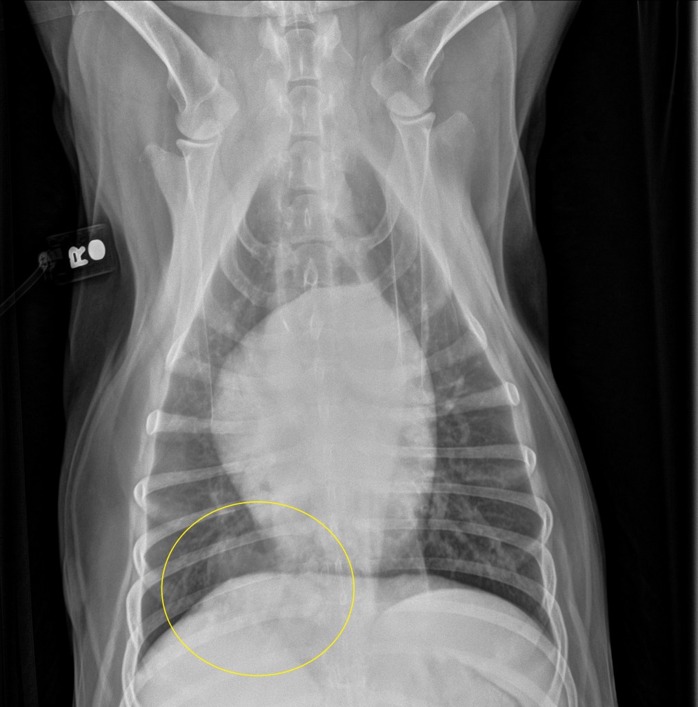
Thoracic radiographs: A mildly ill defined ovoid ~5.0 x 5.3 cm soft tissue opaque mass is located in the dorsal aspect of the right caudal lung lobe in the 7th-10th intercostal spaces. No evidence of intrathoracic lymphadenopathy. Concurrent mild bronchial and interstitial pattern throughout the lungs (likely an age appropriate finding). Findings consistent with primary (e.g. carcinoma) or slightly less likely metastatic or multicentric (e.g. lymphoma) pulmonary neoplasia. Much less likely differentials include granuloma secondary to fungal disease (patient lives in the northeast portion of the country with no travel history) or parasitic infection.
Further diagnostics or treatment: Small chance of ultrasound guided fine needle aspirates of the mass (if an acoustic window can be found to this mass) for possible cytological diagnosis. Alternatively, consultation with an oncologist and/or surgeon could be considered to discuss risk/benefit of excisional biopsy and histopathology of the lung mass.
Pathology of hypertrophic osteopathy: Incomplete understanding. It is often associated with intrathoracic disease/lesions (both infectious and neoplastic) but is rarely associated with neoplasia of the bladder, liver, or ovaries. Reported pathological findings include overgrowth of vascular connective tissues in extremities and subsequent fibroid metaplasia and subperiosteal new bone formation.
